In-Office Treatment of Chronic Sinusitis
The following represents a greater than 2 year longitudinal assessment of a single patient undergoing in office balloon sinus dilation in 2016. In order to effectively demonstrate the clinical outcome, we have summarized the findings below.
Clinical History: Pt presented with a history of right sided facial pain / headache. Over the course of the past year she underwent treatment for migraine without relief. Furthermore, medication aimed at treating nasal congestion and sinus pressure(steroid sprays, antihistamines) was also provided, in addition to antibiotic therapy. Unfortunately, her right sided facial pain / headache persisted despite such efforts. Seeking further options, the patient presented to GNO Snoring and Sinus.
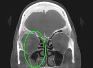
Physical Exam: Clinical and physical exam revealed a component of pressure and discomfort overlying her right maxillary(cheek) and frontal sinus region. There was also a component of right upper dental pain as well. A nasal endoscopy was then performed revealing a normal left sinus drainage pathway, however, the right drainage pathway was noted to have significant swelling – preventing proper ventilation of the sinus cavities. An in-office CT Sinus was then performed (PreOp CT)revealing total blockage of the right sided maxillary/ethmoid/frontal sinus, with mucosal thickening of the sphenoid sinus as well. This resulted in an accurate diagnosis of right sided chronic sinusitis as the cause of her facial pain / headache.
Treatment: The patient underwent in-office balloon sinuplasty of her right maxillary/frontal/sphenoid sinus.
Before:
Preop CT Scan: (2/17/2016)

After:
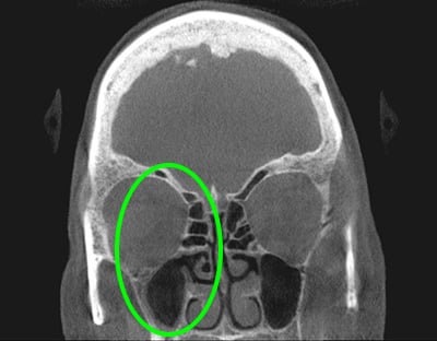
1 Month After: PostOp CT Scan: (4/6/2016)
Summary: Following the in-office sinus dilation, the patient noted a significant decrease in right sided facial pain/headache. The first PostOp CT Scan performed 1 month after the procedure shows improvement in the right sided sinus disease. There is still a level of residual secretion on the CT Scan, and this will continue to drain over the following months.
2 Year PostOp Follow Up: The patient has been followed closely and underwent a CT Sinus in 10/2018 to further assess improvement. As can be seen in the image below there is persistent ventilation and near total clearance of not only the maxillary but also the ethmoid and frontal sinuses on the prior affected right side – demonstrating the long term ventilation provided.
To learn more about in-office sinus dilation and various other office based procedures available at GNO Snoring and Sinus, please contact us to learn more.